Condition
Spondylolisthesis (slipped vertebra)
Spondylolisthesis is the slippage of one vertebra on the one below it. Both common types respond well to physical therapy and injections; fusion is the treatment of choice when they do not.
In plain language
Spondylolisthesis is the slippage of one vertebra on another. There are several types. The degenerative type is commonly seen in older adults, and the "isthmic" type in younger people.
The details
01Symptoms
- Degenerative type (older adults): symptoms of spinal stenosis, such as leg pain with standing or walking, with a greater chance of back pain. If the slip is unstable, back pain becomes more significant.
- Isthmic type (younger adults): the slip is likely present from early adulthood; people tend to develop symptoms in their twenties or thirties.
02Causes
In the degenerative type, arthritis and wear of the discs and joints allow one vertebra to slide forward on the one below. The isthmic type is caused by a small stress fracture in the back part of the vertebra (a pars defect) and is usually present from early adulthood.
03What the MRI shows
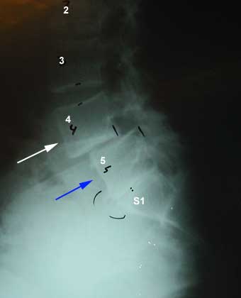
The 4th lumbar vertebra (white arrow) has slipped forward compared with the 5th lumbar vertebra (blue arrow). This is an example of the degenerative type.
X-rays are especially useful for alignment problems like this one: when a vertebra is shifted forward on the one below, that slippage is spondylolisthesis.
04Treatment options
Non-surgical care first
- Physical therapy: both types of slippage respond well.
- Epidural injections can help calm the nerves down.
- Medications during flare-ups.
Surgery
If the pain does not respond to therapy and injections, lumbar fusion is the treatment of choice, with a good prognosis. Fusions done for slippage have more predictable results than fusions done for back pain alone. Fusion is a larger operation, so its risks are discussed in detail first.
05When to see a spine surgeon
- Back or leg pain that is not improving with therapy and injections.
- Leg pain or weakness that is getting worse.
- Any red-flag symptom: see red flags: when not to wait.
Frequently asked questions
01Does a slipped vertebra always need surgery?
No. Both the degenerative and the isthmic types respond well to physical therapy, and epidural injections can help calm the nerves down.
02How well does fusion work for spondylolisthesis?
If therapy and injections do not help, lumbar fusion is the treatment of choice with a good prognosis. Fusions done for slippage have more predictable results than fusions done for back pain alone.
Videos
The anatomy of the low back and the causes of back and leg pain, with MRI findings and common treatments. All videos →
Read transcript: Anatomy of the lumbar spine
Hello, my name is Kambiz Hannani, and today we're going to talk about the bones of the low back or the lumbar anatomy here I have an example of an x-ray looking at the lumbar spine we can see the vertebral bodies the area where the discs are expected as well as the spinous processes we can show that a little bit better on this three-dimensional view of the spine here's the first second third fourth and fifth lumbar vertebrae these are the holes we were talking about where the nerves come out called the foramina and here as we turn the spine over we can see the facet joints which are the joints sitting on the back of the spine and the spinous processes which is the bony elements we feel when we touch our own backs as we turn over to the other side.
We can again see the foramina and finally we can actually see the vertebral bodies from the front on this x-ray we can see the fifth fourth third second first vertebrae the foramina as they come through here as well as the spinous processes back here on the front view of the spine we can see how nicely the spine is straight up and down and we can see the hip bones as they come across.
Although x-rays are helpful MRI is really the diagnostic tool we use to better visualize the soft tissues in the spine here's a fifth fourth third second first lumbar vertebrae these are the discs in between and we can see them pretty nicely here we can see the spinal fluid here as it comes down and protects all the nerves we can see the nerves here this is essentially a normal MRI on the other hand here we have the fifth fourth third second first vertebrae we can see the discs looking pretty reasonably good on these views however the disc between the l4 and 5 looks dark compared to the well hydrated remaining discs this is what we use to determine if there is degeneration of this disc furthermore we can see that the fluid in the spine comes down pretty nicely here until it again hits.
This l45 disc space where a large disc has come out and is really pushing on the nerve structures and the spinal fluid which is likely causing a lot of back and leg pain in this individual x-rays can actually help us a little bit more if you're looking for alignment problems we can see the lumbar spine here nicely aligned however as we get to the sacrum we can see there's a huge shift of the spine pushed forward this whole area should be back up here so that it's a nice continuous element here the other way we can look at the spine is to look at it from the frontal wheel here we can see instead of the spine being straight up and down it's got a large curvature which we call scoliosis when the spine has slipped forward as it has here we call this spondylolisthesis.
We have some x-rays here of a patient who has had surgery by us we can see the disk space with spaces being empty here and then we can see these two disk spaces have a piece of bone in them which is what we have done from the front to try to fuse these segments and then we can see these screws going in and holding the whole structure stable here this is a frontal wheel view again showing us the screws and the bone graft sitting in the middle of the spine.
In summary we have seen the lumbar spine x-ray where things line up really nicely we have seen it when things are not lining up well and we have a spondylolisthesis with the bone falling forward we have seen the frontal view showing us a big curvature which is scoliosis we have seen a very large disc herniation as we can see here coming over and pushing on this nerve as it's being pushed back likely causing significant back and leg pain and we have seen normal discs versus degenerated discs which have lost their luster or the fluid within them and finally we have seen what the spine looks like after instrumentation and effusion has taken place.
This completes our discussion today and thank you for joining us.
A review of the conditions that cause back pain and the different types of surgery used to treat them. All videos →
Read transcript: Types of low back surgery
Hello, my name is Kambiz Hannani, and today I'm gonna talk about lumbar fusions, meaning fusions of the low back region; first things I'm going to look at is looking at the indications meaning the reason for which we do a fusion the most common reasons fusions are done they include slippage of the vertebrae where we can see here the vertebrae all aligned well and then bam there's a forward slippage of this vertebrae relative to the others infections tumors or low back pain indications are important because they determine the success of the surgery the most successful surgeries are done for the slippage infection or tumors low back pain is a bit more unpredictable here we can see a fusion that has been done which screws have been placed on bone graft has been placed in between the vertebrae now there are different ways that a fusion can be performed here we're going to look at the animation to show you exactly what we're talking about this is the lumbar spine the lower back we can see the vertebrae that this spaces which have essentially collapsed because it's a cadaver bone and one way to do a fusion meaning to join these bones together so the bones would not have as much motion or any motion for that matter is to go from the front where we can go in take a piece of disk out put a piece of bone in here and that will allow this bone to fuse between the two bones above and below and prevent this segment from moving another way to do it is to go from the back when we make an incision in the back and we can put some bone on the sides the same idea being the bone can fuse to the two vertebrae and prevent motion at that vertebrae like anything else spinal surgery has risks because these are relatively bigger surgeries risks are a bit greater than some of the other surgeries we're doing the spine like the decompressions the risks include infection bleeding nerve damage including paralysis bowel bladder sexual dysfunction medical complications such as heart attacks pneumonia blood clots or even death although that's not very common there is a chance that a complication may occur symptoms may worsen including back pain or leg pain and there may be a need for additional surgeries nobody can really guarantee the results.
But the success really depends on the indication or the reason for which the surgery is being done here's an example of degenerative disc disease one of the reasons that spinal fusions were performed we can see that these are normal discs and here are the holes through which the nerves come out or the for amen sometimes the dis degenerates as we can see here the disc has degenerated now there is bone on bone arthritis and the forum and where the nerve comes through has collapsed what we do for that or any other type of degeneration for which we do a surgery now one ways to go from the front as we have discussed we go from the belly through the stomach go down take some of that disc out this gives us an area where we can put a piece of bone in so we take a cadaver bone.
Usually from the femur or the thigh bone and we put that right in there and we can see here that bone is sitting in between the vertebrae which allows for these bones to fuse another way to do this is to put some screws into the back to hold things still in this case we are using a minimally invasive way to put the screws in in which we put a cannula down through very small incisions about a centimeter or similar and a half through the skin unlike originally when you use the fusions requiring very big incisions in the back and moving of the muscles which was very painful this allows us to put the screws essentially right through a small incision through the skin all the way down into the spine and then what we need to do is put a rod in between these screws which in.
This case we do by bringing it through an arc as we can see over here right through the screws and that allows us to lock the spine in position and allows the bone to heal up and create a fusion as we can see here where the screws have gone in and have locked into position this is essentially what it would look like with the screws being in place sometimes we take the disc out and put a piece of bone here sometimes we don't and we can see what their final results should look like another way that this can be done is to go ahead and get the disc from the back instead of going through the belly here again we're doing it minimally invasively meaning a small incision is placed.
We can go ahead and put a light source in we can see the bone take off some of the bone from the back this gives us direct access to the disk space which is right here then we can go in take some of the disk out you from the back and then the idea is we like to get in there and expand that disk like we do here where the disc at collapse down by expanding this disk we open the forum and I also bring the disc level back to the way it was before it collapse down this also allows us to put some bone in and after we put the bone into this space we can see the piece of bone we can put some screws and lock it in so this also is another way of doing it doing this fusion and instead of doing a front and back fusion so in summary the most important thing we look at is the indications meaning the reasons for which we were doing a fusion which determines the success rate we really need to be aware of the risks and the benefits involved the risks are clearly present and need to be balanced versus how much improvement you're probably going to be getting and there are different ways of doing it doing this fusion either from the front the back or a combination.
Related procedures & care
Information is general and not a substitute for medical advice.